
Contributor toolkit
Welcome to the Contributor Toolkit. This resource provides healthcare professionals with all necessary forms, diagnostic guidance, and submission instructions for contributing confirmed cases to the IMP Registry.
Please ensure that all relevant documents, if required, are completed and sent to the study team after you receive your login details for case submission. Completed documents should be emailed to the address provided on the IMP Registry database website.

📄 Consent and Information Materials
Before submitting a case, appropriate patient consent or approval must be in place:
-
For UK cases, signed patient consent is required and should be included with the submission.
-
For cases outside the UK, contributors should follow their local institutional approval processes, if applicable.

📝 Data Required for Case Submission
-
Patient demographics
-
Clinical history (obstetric, gynecological, medical, surgical)
-
Ultrasound assessment and other imaging modalities (e.g., MRI) required for IMP diagnosis
-
Imaging files:
- Image 1: Longitudinal view of uterus with IMP
- Image 2: Transverse view of uterus with IMP
- Image 3: Interstitial portion of fallopian tube with IMP
- Additional: 3D volumes and video of imaging (if available) -
hCG measurements (if taken)
-
Data on management and outcomes
-
Data on future fertility, when available

ℹ️ Diagnostic Criteria for IMP
To be accepted into the registry, cases must meet the following diagnostic standards:
-
Implantation of the gestational sac within the myometrium of the uterine corpus, located above the internal cervical os, and clearly separate from the interstitial portions of the fallopian tubes.
-
Implantation occurs beyond the endometrial-myometrial junction.
The following papers may assist you with diagnosis.
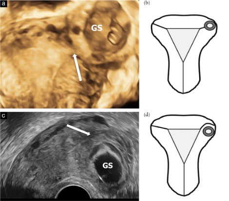
Distinguishing Intramural from Interstitial Pregnancies on Ultrasound:
(a) 3D ultrasound of gestational sac in interstitial portion of fallopian tube (arrow).
(b) Schematic of complete interstitial pregnancy.
(c) 2D ultrasound of gestational sac in anterior myometrium, beneath the interstitial portion (arrow).
(d) Schedmatic of complete intramural pregnancy.
Reproduced from Nijjar et al. (2023), Ultrasound in Obstetrics & Gynecology. Licensed under CC BY-NC-ND 4.0.

❓Need Help?
If you are unsure whether a case qualifies or have questions about the forms or submission process, please contact us: